

Bone pain
Looking for the area of active bone change when pain has not been explained by other tests.
Skeletal Imaging
Bone scintigraphy to show active bone change across the skeleton.
Whole-body bone scans, three-phase scans, bone SPECT-CT and bone marrow imaging to help assess bone pain, injury, infection and spread of disease to the skeleton.
With Dr. Noaline Sinha, Nuclear Medicine & Radiotheranostics.
Artemis Hospital, Sector 51, Gurugram
About Skeletal Imaging
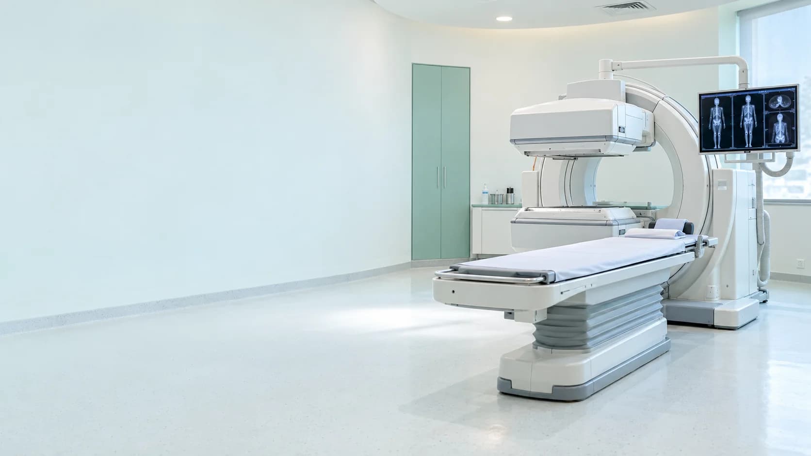
A bone scan uses a tracer that collects in areas where bone is changing. Because this can happen before changes are obvious on X-ray, it can show activity across the whole skeleton in one study. SPECT-CT adds detail about the exact location.
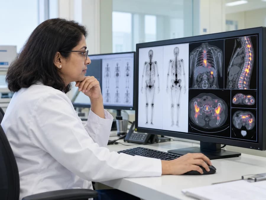
A whole-body scan looks at the entire skeleton, while a three-phase scan adds early images of blood flow to a specific area.
Bone SPECT-CT localises an abnormality precisely, and bone marrow scintigraphy looks at the distribution of marrow.
Your doctor will advise which study is appropriate for your clinical question.
Share your details and our team will get in touch.
Studies Included
Each study is chosen to match the clinical question. This list is provided for patients and for referring clinicians.
4 studies
When It May Help
Your treating doctor recommends imaging based on your symptoms and clinical needs.
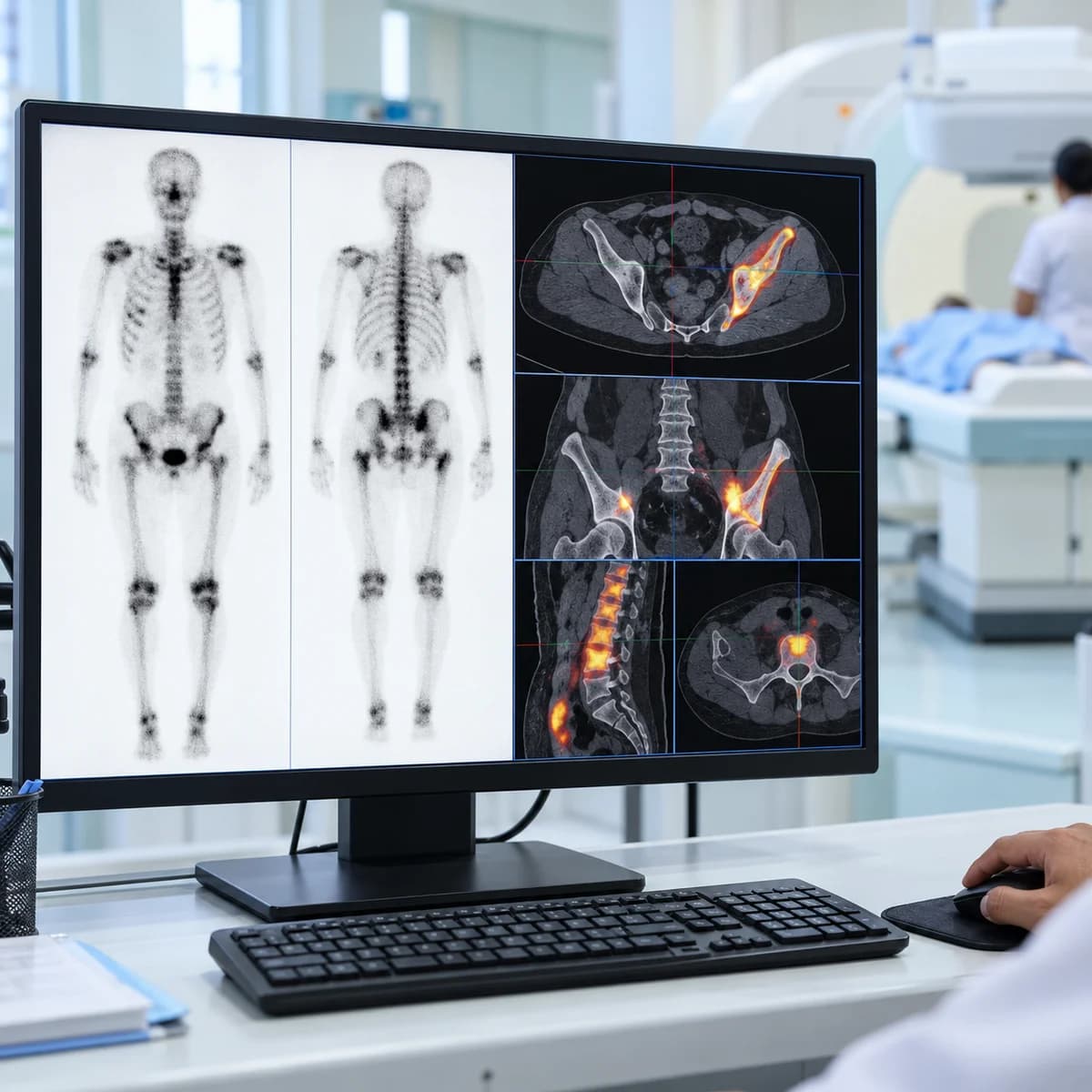
Looking for the area of active bone change when pain has not been explained by other tests.
Checking the skeleton for involvement in people with cancers that can spread to bone.
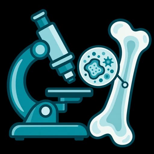
Three-phase imaging can help assess bone and joint infection or inflammation.

Assessing stress injuries, fractures that are hard to see and other bone conditions, as advised by your doctor.

Your Visit, Step by Step
What happens during a gamma camera or SPECT-CT visit.
The team reviews your referral and relevant medical history.
A tracer is administered, followed by a resting period.
You lie on the scanner table while images are acquired.
The team explains the next steps and reporting process.
The exact process depends on your scan and clinical needs.
Before Your Visit
Bring the referral and earlier X-ray, MRI, CT or scan reports where available.
You will usually be asked to drink plenty of fluids after the injection and to empty your bladder before imaging. The team will give you exact instructions.
Whole-body bone scans have a waiting period between the injection and the images. The team will tell you whether you can leave and return.
You may be asked to remove metal objects. Mention recent surgery, fractures or other scans using contrast or tracers.
Let the team know before your appointment.
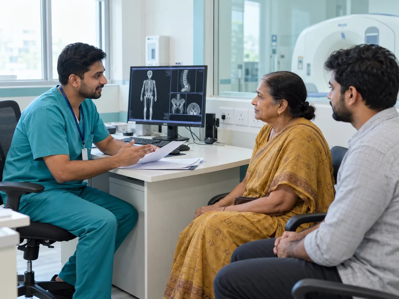
Preparation varies by study. Contact the team if you are unsure about any instruction.

For Referring Clinicians
A short guide for referring doctors.
The clinical question, relevant history, biochemistry or histology where available, and previous imaging reports.
The studies listed on this page are chosen to match the clinical question. If you are unsure which is most appropriate, we are happy to help.
Studies are reported by a nuclear medicine physician, and findings can be discussed with the referring team.
Call or write before referring if you would like to discuss suitability or timing.
Questions before you refer? Contact Dr. Sinha directly.

Your consultant
Chairperson of the Department of Nuclear Medicine & Radiotheranostics, with over 25 years in molecular imaging and radionuclide therapy.
Over 25 years in nuclear medicine, leading the Department of Nuclear Medicine & Radiotheranostics.
Certified Radiation Safety Officer (RSO-II) by the Atomic Energy Regulatory Board.
Works alongside referring and treating clinicians on every case.
Dr. Noaline Sinha
Chairperson, Nuclear Medicine & Radiotheranostics
FAQs
Answers to questions patients often ask before a bone scan.
The tracer takes time to collect in the bones and for the rest to clear from the body, which gives clearer images. The team will explain the timing for your study.
It adds early images taken right after the injection that show blood flow to an area, followed by later images of the bone itself. It is used for specific questions such as infection or injury.
No. A bone scan shows bone activity rather than detailed structure, so it is often used together with X-ray, CT or MRI.
Nuclear medicine studies use a small amount of radioactivity and the team follows radiation safety practices. Tell the team if you are pregnant or breastfeeding, or think you might be.
Your referring doctor will usually discuss the findings with you. Contact the team about when your report will be available.
More Services
Imaging and therapy services offered by the department.
Oncology & Molecular Imaging Neurological PET-CT
 Cardiac PET-CTInfection, Inflammation & Skeletal ImagingCardiac Nuclear MedicineNeurological ImagingRenal & Genitourinary Imaging
 Hepatobiliary & Gastrointestinal Imaging
 Endocrine Imaging
 Pulmonary ImagingInfection, Lymphatic & Other ImagingRadioiodine Therapy
 Peptide Receptor Radionuclide Therapy (PRRT)
 Prostate Cancer Theranostics
 Other Targeted Radionuclide Therapies
 Therapy Planning, Imaging & Dosimetry

Fill in your details and our team will get back to you to confirm your appointment.

Take the Next Step
Reach out to schedule a personalised consultation or speak with our team. We’re here to guide you with clarity, compassion, and expert care — every step of the way.
Schedule a personalised consultation with Dr. Noaline Sinha at your convenience.
Have questions? Our care team is here to help you with guidance and information.
Consultations are held in person in Gurugram. The team will confirm the details with you.